


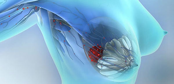
Circulating tumor cells (CTCs) are tumor cells circulating freely in the peripheral blood of patients. The characterization of CTCs is considered as a real-time “liquid biopsy” that provides an ongoing picture of a patient’s cancer status, offering valuable insight into personalized anticancer therapy.
CTCs are very rare and highly heterogeneous, possessing tumor-specific antigenic and genetic characteristics. One of the most commonly used techniques to isolate CTCs is based on the enrichment of tumor cells that express epithelial cell adhesion molecules (EpCAM). This approach, however, can overlook CTC subpopulations that have undergone an epithelial-mesenchymal transition (EMT). It is believed that this EMT process allows the dissemination of CTCs from primary tumors into the circulation. During the EMT process, CTCs lose their epithelial characteristics and acquire more mesenchymal-like phenotypes.
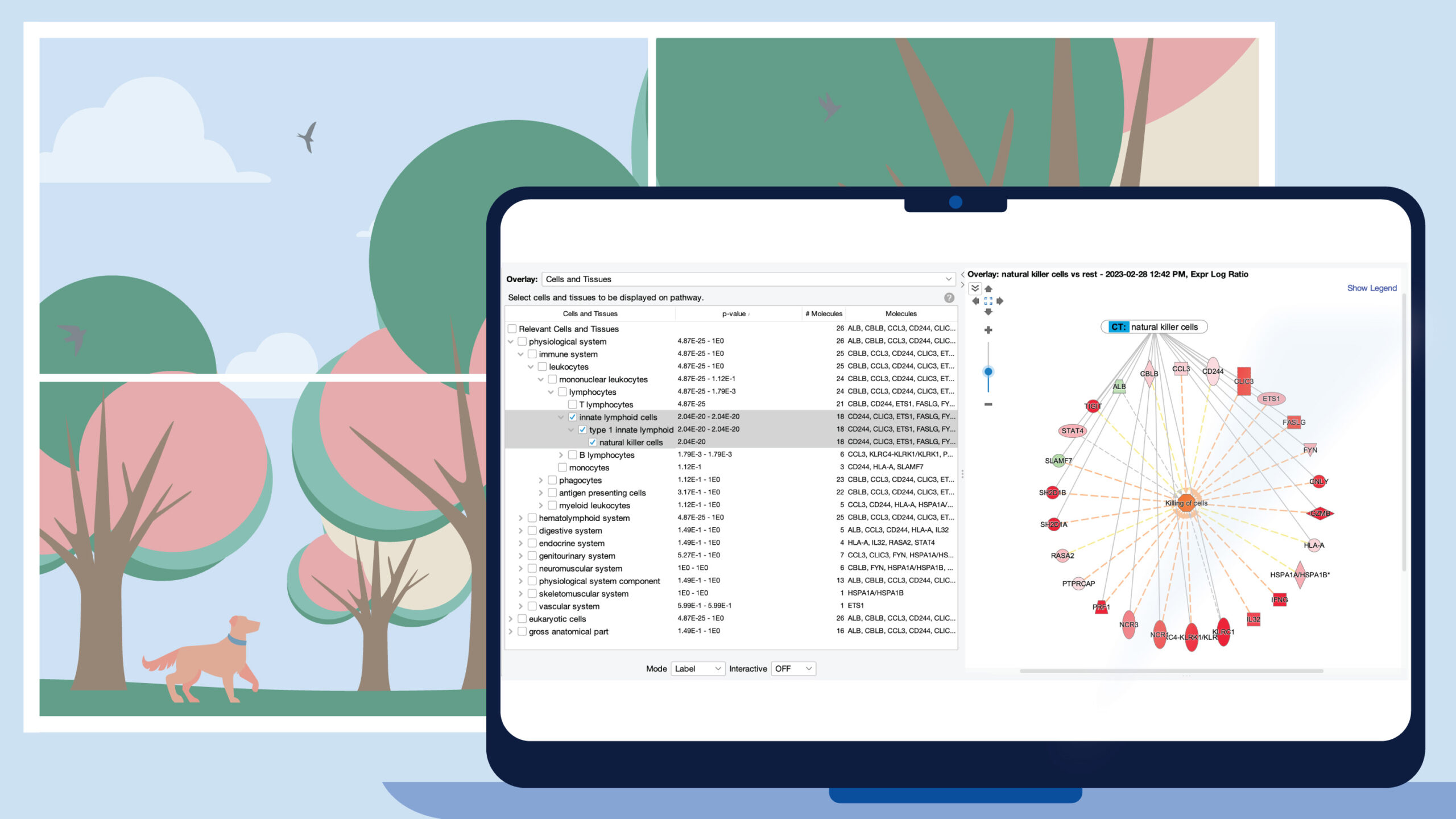
QIAGEN has developed an advanced system that allows enrichment of these cancer cell subtypes from patient blood samples. To accomplish this, we use an antibody-conjugated magnetic bead isolation followed by molecular profiling of captured CTCs. This innovative approach is based on a unique mix of antibodies directed against various tumor cell-associated antigens. By using this technique, you will be able to identify cellular changes in antigen profiles once they develop an EMT or tumor stem cell phenotype, thereby ensuring that your enrichment includes these potentially crucial cells for analysis. This system has been widely used in characterizing cancer progression for targeted therapies.
In a recent investigation from the lab of Dr. Sabine Kasimir-Bauer, Maren Bredemeier and colleagues used the panel AdnaTest EMT-2/Stem Cell Select (QIAGEN Hannover GmbH, Germany) to enrich and profile the expression of 46 genes in CTCs of metastatic breast cancer (MBC) patients. The study was based on 2×5 ml blood from 45 MBC patients and 20 healthy controls, collected at the time of disease progression (T0) and at 2 consecutive clinical stagings (T1 and T2).
One interesting finding is that the multidrug resistant protein gene MRP1 showed a significant difference in expression in overall responders to treatment vs overall nonresponders. In order of significance, VEGFR1, keratin (KRT) 19, EGFR, MET1, ALDH, progesterone receptor (PR), UPA, cathepsin D, KIT1 and Ki67 were differentially expressed in CTCs of patients who had developed liver metastasis as compared to patients without liver metastasis. The patients with liver metastasis showed a significantly lower level of estrogen receptor (ER), PR, HER2, mammaglobin and KRT19 compared to other patients. These preliminary results indicated that CTCs can not only be used as a monitoring tool to guide anticancer therapy, but also allow prediction of the site of metastasis, which may enable a more precise therapeutic decision.
To get more information about this application in MBC research, visit Poster 19, Section 23, at 1 p.m. – 5 p.m., on Sunday, Apr 17, 2016 at AACR in New Orleans.
Find more details about our activities at AACR.